Radiology

All radiological equipment at Charme Clinique is digital
- Ensuring a high image quality
- Enabling significant radiation reduction compared to images taken with the classical plate radiology technique
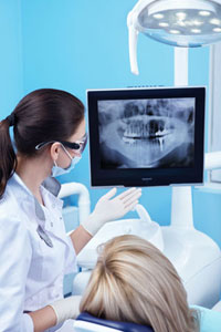
- Displaying test results on a computer in each dental surgery thanks to network software – the specialist dentist may quickly confirm the diagnosis and make a proper treatment decision
- Enabling the results to be recorded on a CD/DVD or external USB memory device.
We perform:
intraoral teeth imaging by a digital radiography system – performed in every surgery room directly during the treatment procedure; the sensor absorbs about 90% of irradiation, making the examination safer than the classical method, by ensuring a minimal dissipation effect;
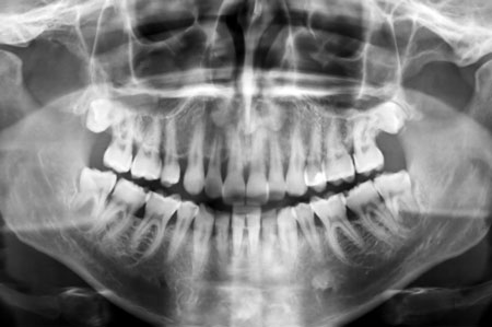
pantographic imaging – overview photos presenting the general condition of all teeth and tissue surrounding jaw bones, lower jaw, jaw joints and maxillary sinuses;

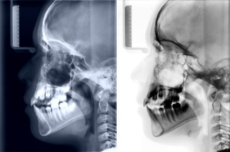
cephalometric imaging – applied most often in orthodontics for face development assessment and planning malocclusion and teeth displacement correction;

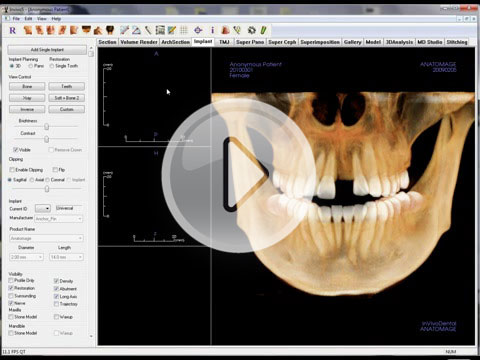
tomographic imaging – 3D photos enable the dentist to see the exact location and size of all anatomic structures and pathologic lesions so as to plan procedures and accurately assess the risk of the planned treatment and the expected effect of procedures. Tomography is used in planning implant placing, dental and jaw surgery, periodontics, orthodontics and advanced endodontics (root canal treatment).


